
Explainer · July 6, 2026 · 6 min · By Tariq Mehmood
Mammograms With Breast Implants: How Screening Actually Changes
Implants do not raise breast cancer risk, but they do change the mammogram itself: extra views, longer appointments, and a technologist who needs to know what she is working with. Here is how to prepare.
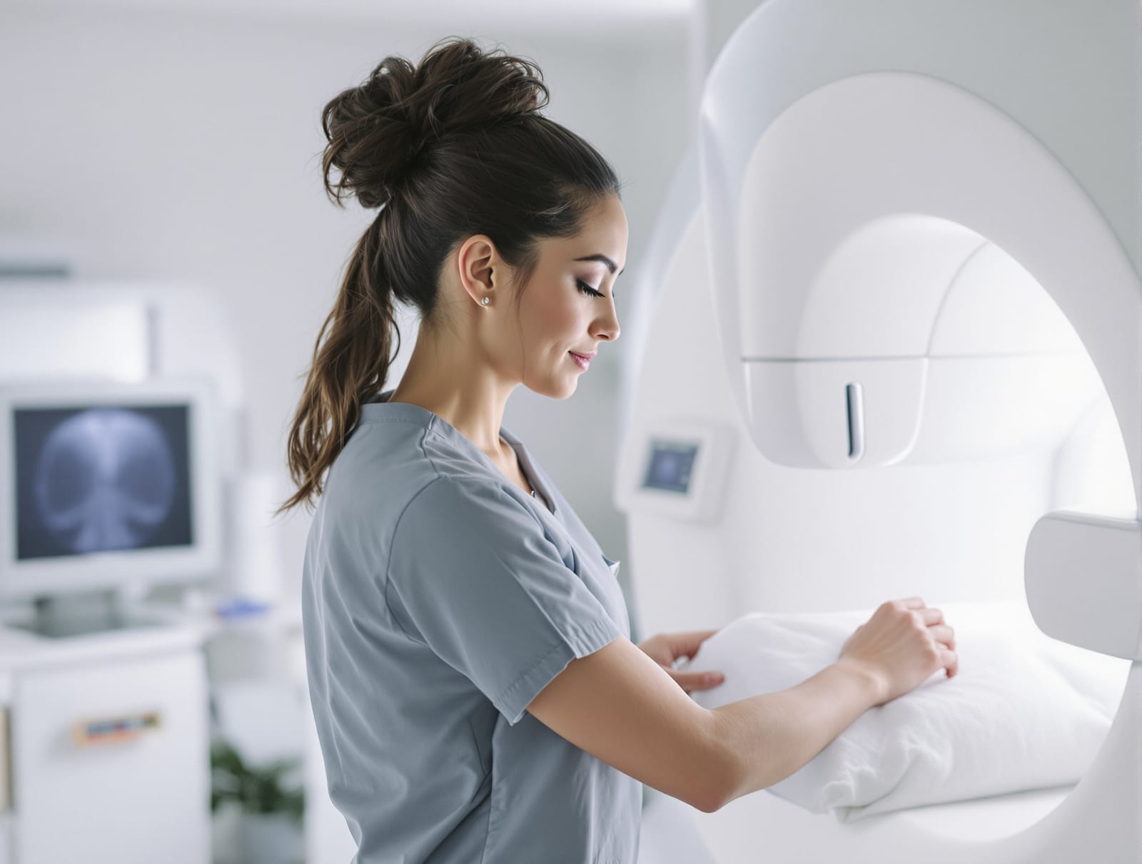
Breast implants change many things about routine health care, and one of the least discussed is the screening mammogram. Every year, augmented patients across Los Angeles arrive for their first mammogram after surgery without knowing the exam will be longer, involve extra images, and require a technologist with specific training. None of that is a reason for alarm. It is a reason to prepare. This explainer covers exactly what changes about breast cancer screening once implants are in place, and the short checklist that makes the appointment go smoothly.
First, the reassurance: implants do not raise breast cancer risk
Decades of study have found no increase in breast cancer risk among women with saline or silicone implants, and the American Cancer Society states plainly that implants do not raise that risk. What implants change is the imaging. Both saline and silicone are radiopaque, meaning X-rays do not pass through them, so an implant appears on a mammogram as a bright white mass that can hide the breast tissue behind and around it. Screening still works with implants. It simply requires a modified technique, and you follow the same screening schedule as any woman your age unless your doctor says otherwise.
The Eklund technique: why you will get extra pictures
A standard screening mammogram takes two images of each breast. For patients with implants, the technologist adds implant displacement views, a method first described by radiologist G. W. Eklund in 1988 that remains the standard today. The technologist gently pushes the implant back toward the chest wall, pulls the natural breast tissue forward onto the imaging plate, and compresses only the tissue. The result is usually four images per breast instead of two: standard views that include the implant, plus displacement views that show as much implant-free tissue as possible. The appointment runs longer, and the positioning takes real skill, which is exactly why the facility you choose matters.
Placement changes what the radiologist can see
Where your implant sits affects how much tissue the displacement views can capture. Implants placed beneath the pectoral muscle allow more tissue visualization on mammography than implants placed above it, because the muscle holds the device back and away from the gland. This is one of the quieter arguments surgeons weigh when recommending submuscular placement for younger patients who have decades of screening ahead of them. Capsular contracture matters here too. A tightly contracted capsule stiffens the breast and makes displacement views harder to perform well, which is one more reason to report new firmness to your surgeon rather than ignore it.
Can the compression rupture an implant?
This is the question patients ask most, and the honest answer is that rupture during a properly performed mammogram is rare. Cases have been reported, and the FDA lists mammography-related rupture as a known but uncommon possibility, but the risk of skipping breast cancer screening is far larger than the risk of the exam. The practical safeguard is disclosure: when the technologist knows implants are present, compression is applied carefully and the displacement technique keeps most of the force off the device entirely. Older implants and implants with known shell concerns deserve a conversation with your surgeon before the appointment, not a skipped exam.
Cancer screening and rupture screening are different exams
A mammogram looks for changes in breast tissue. It is not the tool used to check whether a silicone implant has silently ruptured. That surveillance runs on its own schedule, typically ultrasound or MRI starting five to six years after placement, which we covered in detail in our guide to FDA monitoring recommendations. Some patients coordinate the two, booking rupture surveillance and the annual mammogram in the same season so nothing slips. What you should not do is treat a normal mammogram as proof your implants are intact, or a normal implant ultrasound as proof your tissue is healthy. Each exam answers one question.
How to prepare: a five-item checklist
First, tell the scheduler you have implants when you book, so the facility allots time for the extra views. Second, choose an FDA-certified mammography facility that regularly images augmented patients, and ask that question directly when booking. Third, bring your implant card or at least know your implant type and whether the device sits above or below the muscle, since both details help the technologist plan positioning. Fourth, mention any firmness, pain, or shape change before imaging begins. Fifth, stay consistent: the Mayo Clinic notes that comparing current images against prior ones is central to reading a mammogram, so use the same facility or bring your prior images with you.
What about 3D mammography?
Digital breast tomosynthesis, the 3D mammogram now common across Los Angeles imaging centers, takes multiple thin image slices through the breast and can help radiologists see around overlapping structures. Augmented patients still need displacement views with 3D systems, but many radiologists find the added slices useful in implant cases. If you have dense tissue on top of an implant, ask whether supplemental ultrasound makes sense for you. That is a radiologist's call, made case by case.
The takeaway
Implants and mammograms coexist just fine, provided the imaging team knows what it is working with. Stay on the standard screening schedule for your age, book with a facility experienced in implant displacement views, disclose your implants every time, and keep cancer screening separate in your mind from rupture surveillance. Augmentation is a long-term relationship with your own health care, as we laid out in life with breast implants, and the annual mammogram is one of its simplest, most valuable habits.
Related reading: Implant safety and monitoring: what the FDA actually recommends.